Description
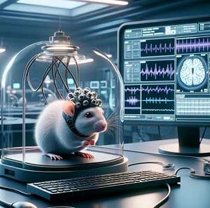
Altogen Biosystems developed rat brain–computer interface with proprietary Altogen® delivery system. System utilize innovative high bandwidth brain chips (a brain machine interfaces to connect animals and computers). Our technology is focused on development of advanced brain-machine interface for animals. Current research prototypes were developed and successfully tested in rats as an animal model of Alzheimer’s disease and Parkinson’s disease, as well as traumatic brain injury and spinal cord injury models. Our proprietary technology create a communication pathway between the rat brain and computer, allowing electrodes that are embedded in the brain, to detect and record rat neural activity and stimulate specific areas of the brain to induce desired responses. We developed proprietary delivery technology with high efficiency effect on targeted parts of the rat brain as well as delivery of biomolecules (plasmid DNA and small RNA) into targeted tissue. Our brain-targeted delivery system utilize nanoparticle-based cargo carriers and optional electroporation application (electroporation is a physical transfection method that utilizes an electrical pulse to create temporary electropores in cellular membranes enabling cargo molecules to be delivered into cells and targeted tissues).

Altogen® delivery system allows advanced real-time testing of novel medicines against Alzheimer’s and Parkinson’s diseases. We have identified highly effective antioxidants in the traumatic brain injury animal model. Our technology of rodent brain-machine interface was successfully tested in multuple rat disease models. System uses rats with electrodes (brain chip) implanted into the brain regions (also called “rat-cyborgs”) that are controled by the brain-machine interface (BMI) technology that seeks to monitor and control a rodent’s brain by an external stimulus and Altogen® biomolecule delivery system. Altogen Biosystems has innovated cutting-edge, high-capacity brain chips, enhancing the interface between animals and computers. This technology centers on the advancement of sophisticated brain-machine interfaces tailored for animals. The current research prototypes have been developed and rigorously tested on rats, serving as an animal model for Alzheimer’s disease, Parkinson’s disease, and spinal cord injury models. Historically, the laboratory rat has made significant contributions to neuroscience research. It has been a favored model for studying Alzheimer’s disease, although its prevalence has waned over the last decade. This decline is attributed to the slower pace of advancements in genetic manipulation techniques for rats compared to mice.

The company’s focus is on developing a functional interface between the rat brain and computer, that is implanted in the brain through a minimally invasive surgical procedure. A brain chip, also known as a neural implant or a brain-computer interface (BCI), is a type of device that is designed to directly interface with the brain in order to manipulate or record its activity. Brain chips are implanted into the brain through a surgical procedure, and they are intended to demonstrate clinically relevant improve for a range of neurological disorders, including Alzheimer’s and Parkinson’s, blindness, and hearing loss. Progress in brain sensing technology is opening up new possibilities for developing neural interfaces that can more efficiently restore or mend neural function and unveil key aspects of neural information processing. However, to fully harness the potential of these bioelectronic devices, we need to comprehend the capabilities of upcoming technologies and identify the most effective approaches to transform these technologies into products and treatments that can enhance the quality of life for patients suffering from neurological and other disorders. The field of artificial intelligence (AI) and robotics is continually expanding its exploration of the junction between living organisms and machines. There is significant focus on AI-driven, motorized prosthetic limbs and brain-machine interfaces that link these prosthetics to human brain activity. The ultimate goal is to perfectly emulate human bodily functions and provide medical aid to those impacted by disease and injury. Yet, to better meld mechanical elements, like prosthetics, with the human body, there’s a need to develop a vast, sophisticated, and flexible sensor that encompasses both the body and the mechanical parts. There are significant developments in the design and miniaturization of implantable devices, often called “brain-computer interfaces” (BCIs). These devices can read brain signals directly, and in some cases, send signals back to the brain. They hold promise for treating a wide range of conditions, from paralysis to mental illness.
What are brain-computer interfaces (BCIs)?
A brain-computer interface (BCI) is a direct communication pathway between the brain and an external device. BCIs are often used in research and are also being developed for medical applications, assistive technologies, and as new ways to interact with computers and other machines.
BCIs work by detecting signals from the brain and converting them into commands for a computer or other device. There are several ways to detect brain signals:
- Non-invasive methods: These involve devices like electroencephalography (EEG) caps that can measure brain activity from the scalp. EEG-based BCIs can be used to create simple commands for a computer, such as moving a cursor on a screen or operating a motorized wheelchair.
- Invasive methods: These involve surgically implanting electrodes into the brain to detect neuronal activity. Invasive BCIs can provide more detailed and accurate readings of brain activity, and can potentially be used to control complex devices like prosthetic limbs.
- Semi-invasive methods: These involve techniques that are partially invasive, such as electrocorticography (ECoG), which entails placing electrodes on the surface of the brain, but not within brain tissue itself.
BCIs have a wide range of potential applications:
- Medical applications: BCIs are being explored as a way to help people with paralysis or other disabilities control prosthetic limbs, communicate, or operate computers.
- Assistive technologies: BCIs can be used to create assistive devices for people with disabilities, such as communication aids for people with severe speech and motor impairments.
- Human-computer interaction: BCIs could potentially be used as a new way to interact with computers, virtual reality environments, and other digital systems.
Developing effective BCIs is a challenging task that requires expertise in neurology, engineering, computer science, and other disciplines. It also raises important ethical issues, such as how to ensure the safety and privacy of users, especially when invasive techniques are used.
What are prosthetic devices (prosthetics)?
Prosthetics, also known as prosthetic devices or prostheses, are artificial body parts that are used to replace body parts that may have been lost due to injury, disease, or congenital conditions. These devices are designed to enhance the function and lifestyle of individuals who have lost limbs or other body parts. Prosthetic devices can replace a wide variety of body parts, from limbs like arms and legs, to smaller parts like fingers and toes. More advanced prosthetics can include parts like artificial eyes, dental prosthetics, or even cosmetic replacements like noses or ears. A prosthesis is often custom-made for the individual to match their needs and body shape, ensuring a good fit and effective functionality. Modern prosthetics can also incorporate technology such as myoelectric sensors that respond to electrical signals from the user’s muscles, or even interfaces that connect to the user’s neural activity, to control the movement of the prosthesis.
How brain activity can be measured and monitored?
There are several types of sensors and techniques used to measure and monitor brain activity. Each of these methods has its strengths and limitations, providing different types of data and requiring different levels of invasiveness.
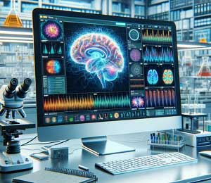
- Electroencephalography (EEG): This technique involves placing electrodes on the scalp to measure electrical activity in the brain. EEG is often used in research, clinical diagnosis, and to monitor the brain during surgery. It provides data with high temporal resolution, meaning it can detect changes over very short periods of time.
- Magnetoencephalography (MEG): This technique measures the tiny magnetic fields generated by brain activity. MEG provides excellent temporal resolution and good spatial resolution.
- Functional Magnetic Resonance Imaging (fMRI): This technique measures changes in blood oxygenation and flow that correspond to brain activity. fMRI provides excellent spatial resolution, meaning it can provide detailed images of which specific brain areas are active, but it has lower temporal resolution compared to EEG or MEG.
- Positron Emission Tomography (PET): PET scans measure the metabolic activity of cells and require the injection of a small amount of radioactive material. They provide good spatial resolution but have lower temporal resolution.
- Near-Infrared Spectroscopy (NIRS): This technique uses infrared light to measure changes in blood oxygenation in the brain. It has moderate spatial and temporal resolution and is less invasive than some other techniques.
- Invasive techniques: In some cases, for example, during neurosurgery or in patients with severe epilepsy, electrodes may be implanted directly into the brain. This provides very precise measurements of electrical activity in the specific areas of the brain where the electrodes are placed.
Each of these techniques measures different aspects of brain activity, and they are often used together to provide a more complete picture of how the brain is functioning. The choice of which technique to use depends on the specific questions being asked, the available resources, and the needs of the patient or research participant.
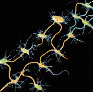
Human brain activity refers to the functioning of the brain as it carries out various tasks and processes. It includes a broad array of physiological occurrences that contribute to how we think, feel, perceive, behave, and interact with the world. The brain contains billions of neurons, which communicate with each other through electrical signals and chemical neurotransmitters. When neurons are activated, they produce an electrical impulse known as an action potential, which travels down the neuron and leads to the release of neurotransmitters. These neurotransmitters cross the synaptic gap between neurons and bind to receptors on a neighboring neuron, potentially leading to a new action potential in this second neuron. This is the fundamental process of information transfer in the brain.
What are rat cyborgs?
A rat cyborg, also known as a robo-rat or ratbot, refers to a rat that has been implanted with electronic or robotic components, allowing it to be remotely controlled or influenced. The goal of such experiments is typically to better understand how the brain works, to develop more advanced neuroprosthetics, or to explore new possibilities for human-machine interaction. The concept of a rat cyborg is controversial due to ethical concerns regarding animal welfare. However, it also offers potentially valuable insights into the neurology of mammalian brains, including our own. It’s a testament to the rapidly advancing field of neuroscience and its intersection with technology, but it also underscores the critical need for ethical considerations in such research. For instance, researchers have implanted electrodes into the brains of rats and can transmit signals that instruct the rat to move in certain directions. This technology could potentially be used in future applications such as search and rescue operations, where rat cyborgs might be used to navigate tight spaces or dangerous areas where it would be unsafe for humans.


