
Description
Purchase Orders: Click “Add to Cart” button to order, then email PO to orders@altogen.com.
Product Availability: In Stock.
Transfection Reagent for Keratinocyte Cells (Keratinocytes)
-
Two component formulation enhances lipid mediated transfection efficiency
-
Optimized easy-to-use transfection protocol provided for transfection of siRNA, DNA, mRNA, and microRNA
-
Kit includes Transfection Enhancer reagent and recommended transfection protocol
-
High transfection efficacy in the presence of serum
-
Expand your RNAi application with a reagent optimized for delivery of both siRNA and plasmid
-
Reproducible transfection results
-
Works well for standard reverse transfection and high-throughput applications
-
Download in vitro Keratinocyte transfection protocol: [PDF]
- Download Keratinocyte CRISPR/Cas9 transfection protocol: [PDF]
-
Download PowerPoint presentation for Keratinocyte cells transfection kit: [PPT]
- UPC/GTIN/EAN: 860002089781
-
Brand: ALTOGEN®, developed and manufactured by Altogen Biosystems
Transfection Efficiency:
Reagent exhibits at least 85% transfection efficiency of siRNA delivery. Transfection efficiency was determined by qRT-PCR.
Product Description:
Enhanced transfection kit designed for superior efficiency for keratinocyte cells, both cell lines and primary keratinocytes. Kit includes transfection reagent, complex condenser, and transfection enhancer reagent.
Transfection Protocol and SDS:
Download Altogen Biosystems Keratinocyte Transfection Protocol: [PDF]
Download SDS: [PDF]
Keratinocyte Cell Line:
Research using keratinocyte cells has contributed significantly to our understanding of skin biology and the development of skin diseases. For example, studies have shown that exposure to ultraviolet radiation can induce DNA damage and promote the development of skin cancer through the activation of certain signaling pathways in keratinocyte cells. Additionally, keratinocyte cells have been used to test the efficacy of various therapeutic agents for skin diseases, including topical medications and gene therapy approaches. Keratinocytes are the most prevalent type of skin cells, comprising nearly 95% of the epidermis. Keratinocyte cells originate in the basal skin layer and grow densely outward through the layers of the epidermis. Keratinocyte cells are continuously shed and replaced from the outer layer. They are considered the building blocks of skin because they are responsible for keratin production, the key component of hair and nails. Keratinocyte cells are tightly interwoven and form seams between the nerves and underlying tissues of the epidermis. This allows them to create a protective barrier to prevent infectious substances coming into the body through the skin. Also, keratinocytes play an essential role in wound healing, demonstrating a higher expression of PDGF A (fibroblast stimulating platelet-derived growth factor) than fibroblasts and regulating fibroblast activity.
Keratinocytes are commonly used to develop in vitro techniques to assess skin irritants. The cells are also the principal source of the proinflammatory cytokine IL-1 in the epidermis. Studies have found that Bcl-2 overexpression protects keratinocytes from induced apoptosis. Keratinocytes are sometimes divided into smaller cell types, such as basal keratinocytes, which are located near the basal layer. There are multiple biomedical research applications using keratinocytes. Altogen Biosystems offers advanced cationic lipid-based Keratinocyte transfection reagent kits that can efficiently aid biomedical research related to keratinocytes. These kits can be utilized for the development of stable keratinocyte cell lines and yield high transfection efficiency, as detected by qRT-PCR. Keratinocyte cells are of great interest in research due to their role in maintaining skin health and their involvement in various skin diseases. For example, studies have shown that defects in keratinocyte differentiation and proliferation can contribute to the development of skin disorders such as psoriasis, atopic dermatitis, and skin cancer. Keratinocyte cells can be isolated from skin biopsies or obtained from commercial sources. They can be grown in culture and used as a model system for studying various aspects of skin biology, including the mechanisms underlying keratinocyte differentiation and proliferation, wound healing, and the response of skin cells to environmental stressors.
Data:
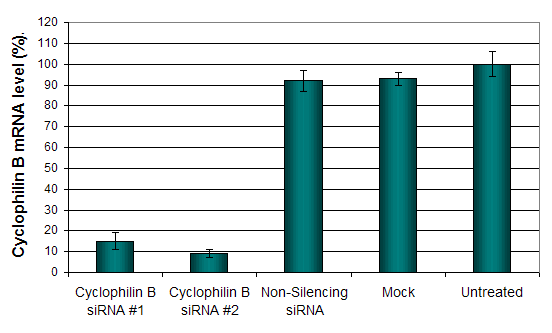
Figure 1. Cyclophilin B silencing efficiency was determined by qRT-PCR in the cells transfected by Cyclophilin B siRNA or non-silencing siRNA control following the recommended transfection protocol. Cyclophilin mRNA expression levels were measured 48 hours post-transfection. 18S rRNA levels were used to normalize the Cyclophilin B data. Values are normalized to untreated sample. Data are presented as means ± SD (n=3).
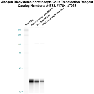
Figure 2. Protein expression of Cyclophilin B in Keratinocyte cells. DNA plasmid expressing Cyclophilin B or siRNA targeting Cyclophilin B were transfected into Keratinocyte cells following Altogen Biosystems transfection protocol. At 72 hours post-transfection the cells were analyzed by Western Blot for protein expression levels (normalized by total protein, 10 µg of total protein loaded per each well). Untreated cells used as a negative control.
Selected in vivo transfection product citations (ALTOGEN® IN VIVO Transfection Kits used in the following publications):
- Nature. 2008 454(7203):523-7. Innate immunity induced by composition-dependent RIG-I …Saito et al [PDF]
- Am J Pathology. 2010 177(4):1870-80. Role of ocular complement factor H in a murine model … Lyzogubov et al [PDF]
- Nature Biotechnology. 2011 29(4):341-5. Delivery of siRNA to the mouse brain by … Alvarez-Erviti et al [PDF]
- Cancer Research. 2011 71(15):5144-53. Inhibition of miR-193a expression by… Iliopoulos et al [PDF]
- RNA. 2010 16(11):2108-19. RNase L releases a small RNA from HCV RNA that refolds … Malathi et al [PDF]
- Diabetologia. 2012 55(7):2069-79. The p47phox- and NADPH oxidase organiser 1 … Youn et al [PDF]
- British Journal of Cancer. 2012 107(3):516-26. TIGAR induces p53-mediated cell-cycle … Madan et al [PDF]
- Hypertension. 2014 63(2):353-61. Tissue transglutaminase contributes to … Liu et al [PDF]
- Circulation Research. 2010 15;107(8). Kruppel-like factor-4 transcriptionally regulates … Cowan et al [PDF]
- PLoS Pathogens. 2012 8(8) Uridine composition of the poly-U/UC tract of HCV RNA … Schnell et al [PDF]
- J Proteome Res. 2012(11) Retinal proteome analysis in a mouse model of oxygen-induced … Kim et al [PDF]
- PLoS Pathog. 2014 10(10) Exosomes from hepatitis C infected patients transmit HCV … Bukong et al [PDF]
Altogen Biosystems is a life sciences company that offers cell type-specific and pre-optimized transfection products, elecroporation kits, and in vivo delivery reagents. Advanced formulation of reagents and optimized transfection protocols provide efficient intracellular delivery of protein, DNA, mRNA, shRNA and siRNA molecules. Read more about transfection technology at Altogen’s Transfection Resource. Altogen Labs provides safety and efficacy preclinical research services. GLP-compliant studies for IND applications, and drug development, including over 90 in-house validated xenograft models, safety toxicology, etc (visit AltogenLabs.com).
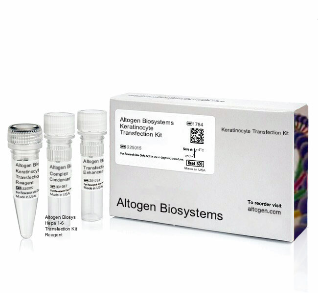
Volume Options:
- 0.5 ml (Catalog #1783)
- 1.5 ml (Catalog #1784)
- 1.5 ml CRISPR (Catalog #2164)
- 8.0 ml (Catalog #7053)
Purchase Orders: Click “Add to Cart” button to order, then email PO to orders@altogen.com.
Product Availability: In Stock.